What is CBCT?
Dental CBCT scanners can be a bit like the marmite of the dental imaging world; loved by some, not by others. Here's what you need to know about CBCT.
What is CBCT / Cone Beam CT?
Dental cone beam computed tomography (CT) is a special type of x-ray machine that allows for more detailed imaging in the diagnosis and case planning process due to its ability to produce three dimensional (3D) images of dental structures, soft tissues, nerve paths and bone in the craniofacial region in one single scan.
CBCT scans let you effectively peel back layers and see your patients anatomy from different angles, and often, uncover areas that need attention which could not have been seen by traditional Orthopantomogram (OPG).
How does CBCT Work?
With Cone Beam CT, an x-ray beam in the shape of a cone is moved around the patient to produce a large number of images, also called views, in order to create a navigable 3D rendering of the patient's dental structures.
The approach of taking multiple exposures allows for more detailed analysis, particularly in regions where a traditional 2D X-Ray would struggle to give accurate imaging.
What does a CBCT image look like?
CBCT scans produce rich 3D images, that can be used to demonstrate key areas to the patient and, since the images they produce are very easy for patients to understand, they often lead to much higher case acceptance rates than traditional 2D scans.
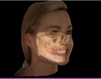
Image courtesy of Carestream Dental
Are CBCT machines hard to use?
Despite producing beautiful, actionable 3D scans that are incredibly complex for computers to assemble, the actual process of using a CBCT is incredibly simple for Dentists.
In terms of managing patient flow, the image capture stage is virtually identical to taking an OPG image - though the patient should always be informed of additional radiation exposure, and consent to it as per your regular procedures.
Do patients appreciate CBCT scans?
The biggest difference in terms of patient flow comes after the scan. Where an OPG image may be used to point at a problem, the 3D scan created by a CBCT allows the dentist to orientate the patient first. Once the patient understands what they are looking at, you can then find the best angle from which to explain the diagnosis and the impact on any surrounding anatomy.
This workflow can be very collaborative, and many dentists find that patients gain a much deeper understanding of their oral health as a result of it. Further, understanding builds appreciation, which is why 3D scans can often contribute to much higher patient acceptance rates than 2D X-rays, even panoramic ones.
What is dental CBCT best for?
While there are countless use cases for dental Cone Beam CT, the most common uses are in endodontics and implant planning.
In the case of endodontics, CBCT can reveal much more depth and diagnostic confidence than a typical periapical (PA) X-ray, and thus be used to make better treatment decisions.
For implant planning, dental CBCT gives a much clearer understanding of the patients bone structure. Where OPG will show the placement of bone structures, CBCT will give a clearer view of factors like bone thicknesses and density, which allows you to better prepare yourself and your patient for the treatment plan.